Project: EQC2021-007761-P
Spanish Ministry of Economy and Competitiveness
Proteomics is one of the omics technologies whose application is growing exponentially. Protein analysis by mass spectrometry is making it possible to explore the proteome of living organisms in depth. Its application to both biological and biomedical problems makes it possible to identify and quantify changes in protein profiles associated with biological and pathological processes. At the level of global proteome analysis, it is a powerful tool. This is why proteomics is often used in discovery stages of research projects by analysing thousands of proteins in a relatively small number of samples. These analyses are really effective in focusing attention on the most relevant proteins involved in the biological systems under study. Although genomic studies are of undoubted value, it is often not possible to link them to function because the transcriptome does not directly correlate with the proteome. On the other hand, complex post-transcriptional regulation means that proteomes often respond to very precise cellular demands, frequently associated with rapid transitions and/or responses over time. All this, together with the cellular heterogeneity that is being revealed in recent years, makes it absolutely necessary to be able to study the proteome with the greatest possible sensitivity.
In order to carry out most proteomic analyses until now, a considerable amount of sample was necessary. The sensitivity of the technique has improved considerably in recent years, from using several million cells to several thousand cells in experiments. Despite this improvement, this global proteomics does not allow us to gain insight into the complex biological processes that originate in or are modulated by interactions between individual cells. There is a cellular heterogeneity, even within each cell type, that could explain many biological and pathological processes that we cannot access with global proteomics. The role of the cellular microenvironment within a tissue in cell physiology and the modulation of responses associated with different pathologies is becoming increasingly clear. Therefore, in order to understand in depth many of the molecular and cellular processes of interest in biomedicine, it is not only important to know which cells are present but also where and with whom they are present, as well as the interactions between them. This information is inaccessible to conventional proteomics techniques, on a global scale, in most cases.
The genomic analysis of single cells, or a small number of them, is opening up a world of new possibilities in biomedical research. At the protein level, confocal microscopy has allowed the study of individual cells, but only of a very small number of proteins and in a targeted manner. In the absence of a methodology equivalent to PCR, access to proteomic analysis of individual cells depends on increased instrumental sensitivity and improved sample preparation processes. In this regard, recent technical advances are achieving unprecedented levels of sensitivity that make it possible to meet the challenge of single cell proteomics. Cutting-edge instruments would make it possible to address a whole series of biological problems in which cellular heterogeneity and its interactions play a relevant role.
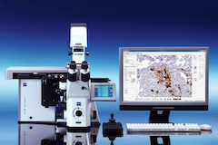
We know that there are many cell types, some of them well characterised. However, despite the morphological differences between cells, we do not always know what makes them different. For example, we know that several types of neurons coexist in each region of the brain. At the morphological level they can be differentiated, but it is difficult to know what their molecular differences are to understand their biology. Frequently, primary cell cultures are used. However, it is not always possible to maintain the original cellular properties in culture so that, in many cases, we cannot be sure that what we analyse in culture is a true reflection of the cell found in the tissue. Complete tissue analysis does not allow us to identify the individual characteristics of each cell type. The equipment acquired in this project, on the other hand, does allow the proteomic characterisation of each cell type. In this situation, given that there are appreciable morphological differences, it is possible to use a laser microdissector to extract the cells of interest from a tissue section and subject them to single-cell proteomic analysis.
Single cell proteomic analysis is a truly innovative technique. Moving from the analysis of thousands of cells, with the associated cellular heterogeneity, to the analysis of single cells is a major technological challenge. However, it is a challenge worth taking on because of the great rewards that lie behind it. The potential for biomedical and biotechnological research is enormous. The performance of recently developed instruments makes it possible to specifically extract the cells of interest from a tissue and carry out their proteomic characterisation. This technology will undoubtedly make a decisive contribution to the advancement of biomedical research.
In this project, we have obtained funding to implement a single cell proteomics platform to be installed in the Proteomics Section of the Central Research Support Services (SCSIE) of the University of Valencia.